
Diabetic Eye Exams in Texas
Protect your vision with specialized diabetic eye examinations designed to detect and monitor diabetes-related eye complications early. Our comprehensive approach combines advanced diagnostic technology with personalized care to preserve your eye health and prevent vision loss.
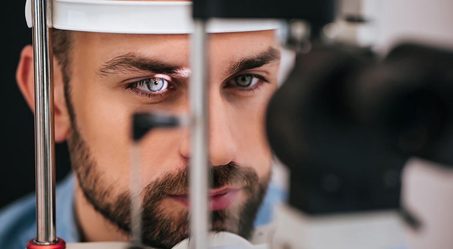
What is a Diabetic Eye Exam?
A diabetic eye exam is a specialized comprehensive eye examination specifically designed for individuals living with diabetes, whether type 1, type 2, or gestational diabetes. These examinations go beyond standard vision testing to focus on detecting and monitoring the unique eye complications that diabetes can cause, including diabetic retinopathy, diabetic macular edema, cataracts, and glaucoma. The exam utilizes advanced diagnostic imaging and careful evaluation of the retina, blood vessels, and other delicate structures of the eye that are particularly vulnerable to damage from elevated blood sugar levels.
Diabetes affects the small blood vessels throughout the body, and the eyes are especially susceptible to this damage. High blood glucose levels can cause the blood vessels in the retina to leak fluid or bleed, leading to vision problems and potential blindness if left undetected and untreated. Diabetic retinopathy is currently the leading cause of blindness among working-age adults in the United States, yet up to 95% of severe vision loss from diabetes is preventable with early detection and timely treatment. Regular diabetic eye exams are essential because eye damage often occurs before any noticeable vision changes, making these screenings crucial for catching problems in their earliest, most treatable stages.
The American Diabetes Association recommends that people with type 1 diabetes have their first dilated eye exam within five years of diagnosis, while those with type 2 diabetes should have an exam immediately upon diagnosis since the disease may have been present for years before detection. Pregnant women with diabetes should be examined during the first trimester and monitored closely throughout pregnancy and postpartum. These regular examinations are not just recommended—they are essential preventive care that can mean the difference between maintaining healthy vision and experiencing irreversible vision loss.
Signs You Need a Diabetic Eye Exam
- You have been diagnosed with type 1, type 2, or gestational diabetes
- It has been more than 12 months since your last comprehensive eye exam
- You are experiencing blurry or fluctuating vision, even if it comes and goes
- You notice floaters, dark spots, or shadows in your field of vision
- You have difficulty with color perception or night vision
- Your blood sugar levels have been poorly controlled or fluctuating
- You are pregnant and have pre-existing or gestational diabetes
Our Approach to Diabetic Eye Exams
Comprehensive Assessment
We begin with a thorough review of your diabetes management, including your HbA1c levels, blood sugar control history, and any previous eye-related concerns. This comprehensive intake helps us understand your individual risk factors and tailor the examination to your specific needs.
Advanced Diagnostic Testing
Using state-of-the-art technology including dilated fundus examination, optical coherence tomography (OCT), and retinal photography, we capture detailed images of your retina and optic nerve to detect even the earliest signs of diabetic eye disease before symptoms appear.
Personalized Monitoring Plan
Based on your examination results and risk profile, we develop a customized monitoring schedule and coordinate with your diabetes care team to ensure comprehensive management. If any concerns are detected, we provide prompt referrals and treatment recommendations.
Specialized Diagnostic Technologies
Our diabetic eye examinations utilize multiple advanced technologies to provide the most comprehensive assessment possible. Optical Coherence Tomography (OCT) creates high-resolution cross-sectional images of the retina, allowing us to detect subtle changes in retinal thickness and identify diabetic macular edema in its earliest stages. Digital retinal photography documents the appearance of your retina over time, enabling us to track even minute changes that might indicate progression of diabetic retinopathy.
The dilated fundus examination remains the gold standard for comprehensive retinal evaluation. By temporarily widening your pupils with eye drops, we can examine the entire retina, including the peripheral areas where early diabetic changes often first appear. We carefully inspect the blood vessels for signs of microaneurysms, hemorrhages, exudates, and neovascularization—all indicators of diabetic retinopathy at various stages.
When clinically indicated, we may perform fluorescein angiography, a specialized imaging technique that uses an injectable dye to highlight blood flow patterns in the retina and identify areas of leakage or poor circulation. We also conduct tonometry to measure intraocular pressure, as diabetes increases the risk of glaucoma, and perform visual field testing to detect any peripheral vision loss that may indicate advanced diabetic eye disease or concurrent glaucoma.
Stages of Diabetic Retinopathy We Monitor
Mild Nonproliferative Retinopathy
Early stage with microaneurysms—small areas of balloon-like swelling in the retina's tiny blood vessels.
Moderate Nonproliferative Retinopathy
Blood vessels that nourish the retina become blocked, with more severe changes visible during examination.
Severe Nonproliferative Retinopathy
Many more blood vessels are blocked, depriving blood supply to areas of the retina and triggering growth factor signals.
Proliferative Diabetic Retinopathy
Advanced stage where new, abnormal blood vessels grow, which are fragile and can leak, causing severe vision loss and requiring immediate treatment.
What to Expect
First Visit (90-120 minutes)
Your initial diabetic eye exam begins with a detailed health history, including questions about your diabetes diagnosis, current medications, blood sugar control, and any vision concerns. We will perform vision testing, measure eye pressure, and administer dilating drops. After allowing 20-30 minutes for full dilation, we will conduct a comprehensive retinal examination and capture diagnostic images. You should plan to have someone drive you home as your vision will be blurry and light-sensitive for several hours after dilation.
Results Discussion (Same Day)
Before you leave, we will review all findings with you in detail, showing you images of your retina and explaining what we observed. If no diabetic eye disease is detected, we will establish a monitoring schedule appropriate for your risk level—typically annual exams for those with well-controlled diabetes and no retinopathy. If we detect any changes, we will discuss the severity, treatment options, and follow-up timeline.
Follow-Up Schedule (Personalized)
Your follow-up schedule depends on your examination findings. Those with no retinopathy and well-controlled diabetes typically return annually. Mild nonproliferative retinopathy may require monitoring every 6-12 months. More advanced changes necessitate more frequent examinations, potentially every 2-4 months, and may require referral to a retina specialist for treatment such as laser therapy or intravitreal injections.
Long-Term Partnership
Diabetic eye care is a lifelong commitment, and we are here to partner with you throughout your journey. We communicate with your primary care physician and endocrinologist to coordinate your overall diabetes management, as blood sugar control directly impacts eye health. We celebrate stable results with you and provide support and rapid intervention if changes occur. Our goal is to help you preserve your vision for life through vigilant monitoring and timely care.
Common Questions About Diabetic Eye Exams
How often should I have a diabetic eye exam?
Will my eyes be dilated, and how long does it last?
Can diabetic eye disease be treated if it is detected?
My vision seems fine. Do I still need a diabetic eye exam?
Does good diabetes control really make a difference for my eyes?
Related Services
Comprehensive Eye Exams
Complete vision and eye health evaluations for all ages, including refraction, ocular health screening, and disease detection.
Glaucoma Screening
Early detection and monitoring of glaucoma through pressure testing, optic nerve evaluation, and visual field assessment.
Retinal Imaging
Advanced diagnostic imaging technology to capture detailed images of your retina for comprehensive eye disease detection and monitoring.
Ready to Protect Your Vision with a Diabetic Eye Exam?
Early detection is key to preventing diabetes-related vision loss. Schedule your comprehensive diabetic eye exam today and take control of your eye health.
Schedule Your Exam